In school laboratory, when tissues are seen under microscope, cells always appear to possess dark coloured nuclei. Why is it so?
1 Answer

May 19, 2017
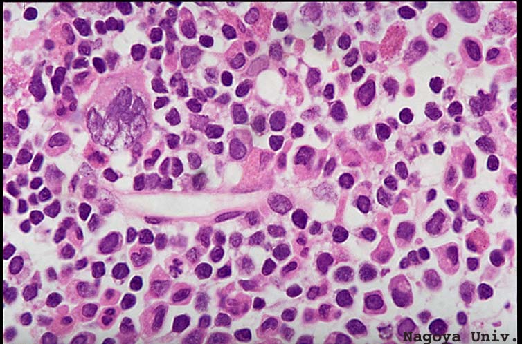
When basic stains are used to prepare tissues for studying under microscope, nucleic acid of nuclei take up more stain and thus nuclei appear dark in colour.
Explanation:
Generally hematoxylin is used to stain nucleus: it is a basic stain and it binds to acidic, negatively charged DNA. Nucleus takes up dark blue to violet colour.
Another stain is used to provide contrasting colour to surrounding cytoplasm of the cell. This is eosin, an acidic stain which provides light pink colour to cytoplasm, by binding with basic amino acids like lysine and arginine.
Histochemical staining technique is an important branch of biology.