What are the horns of gray matter?
1 Answer
The lighter part of the transversal cross section of the spinal cord.
Explanation:
First of all, it is important to say that the position of the white and grey matter is different in the brain from that in the spine.
In the brain, in a sagittal and coronal (=frontal) plane, the outermost part is grey and the innermost is white matter. The reverse is in the spine (however, the plane has to be either coronal (hardly ever used), sagittal (hardly ever used) or transversal (mostly used)) where the grey part is the innermost and the outermost is the white part.
According to your question and referring to the information above, I can freely conclude that this is about the spine.
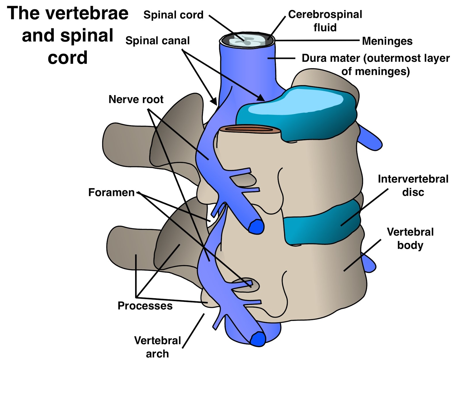
Anatomically, we are informed that the spine is positioned on our back and that it protrudes from the brain until the pelvis. It is protected by vertebrae.
Physiologically, we are informed that its function is to transmit signals from our peripheral nerves via the spinal cord (=spine) to the brain.
Histologically, the anatomical, as well as physiological relations between peripheral and central nervous system, are established.
Take a look at the picture below. (Dorsal = back, posterior; ventral = front, anterior)
Here, you can see two different regions; the darker one and the lighter one. The darker one is the white matter, whilst the darker one is the grey matter.
It is important to say that in the white matter we find dendrites (cell bodies of neurons) and in the grey matter we find axons. This is the same for both the brain and spinal cord.
The lighter part (from now on in the following text: the grey matter) is also known as "butterfly" because it resembles of a butterfly's wings.
As I have said previously, the spinal cord transfers signal from peripheral nerves to the brain. But one more thing it does is known as a reflex. In some textbooks, you will find that the reflex is a consciously uncontrolled reaction, meaning that we do not (have) time to think to respond to an environmental stimulus. But imagine a situation that you are preparing a meal and suddenly you get burned. Would not you feel the pain? Is not the parietal lobe of the cerebrum assigned for sensation? Well, the thing is that during that reflex the signal actually goes to the brain, but perhaps it is a couple of mili or even micro seconds late in comparison to the one of the reflex.
At the top of the picture, you can see that there is even lighter, opening-like area. That area is connected to a sensory (=afferent) nerve. This nerve takes signals, for instance, your hand, and then it sends it to the spinal cord or to be more precise the grey matter of the spinal cord. Once it enters the grey matter it connects to an interneuron which is just a neuron that takes the signal and then transmits it to a motor (=efferent neuron) posteriorly. This is bilateral (on the both sides divided (not fully separated!) by the central canal).
The central canal (=ependymal canal) is a longitudinal canal filled with cerebrospinal fluid (CSF) that connects the cerebroventricular system and the spine.
In a macro view, you can see at what points the sensory and motor nerves enter and exit, respectively.
You might now be asking: if the brain's white and grey matter is differently positioned from the one of the spinal cord, where is the place the change occur?
This is to be double-checked: It could be the in the foramen magnum, where the spinal cord enters the neurocranial skull part.
